Thursday, January 21, 2016

Determinació de: llet, caseïna, altres proteïnes, midó i glúcids reductors i presència de lípids

Determinació de la composició de la llet:
La llet conté vitamines (tiamina, riboflavina, àcid pantotènic i vitamines A, D i K), minerals (calci, potassi, sodi, fòsfor i metalls en petites quantitats), proeïnes, glúcids i lípids. Els únics elements importants que no te són ferro i vitamina C.

Pràctica: identificar tots aquests components (resum de totes les proves d'identificació)

1- Determinació de la caseïna:
La caseïna és una proteïna conjugada del tipus fosfoproteïna que se separa de la llet per acidificació i forma una massa blanca. Les fosfoproteïnes són un grup de proteïnes que es troben químicament unides a àcid fosfòric, en el cas de la caseïna aquests grups fòsfor es troben units als aminoàcids serina i treonina. La caseïna representa entre 77 i el 82% de les proteïnes presents a la llet. Aquesta proteïna presenta una baixa solubilitat a pH 4,6. En la primera part del experiment aïllareu la caseïna.

1- Afegiu 200ml de llet en un vas de precipitats i escalfeu fins 40 graus aproximadament.
2- Traieu-lo del foc i afegiu gota a gota àcid acètic (1ml d'àcid acètic glacial en 10 ml d'aigua destil.lada) amb un comptagotes. Agiteu amb una vareta de vidre fins que acabi de precipitar tota la caseïna. (No afegir massa àcid acètic)

3- Separeu la caseïna amb ajuda d'una espàtula i poseu-la en un vidre de rellotge. Poseu-lo a assecar en la placa calenta.
4- Afegir immediatament en el líquid que us ha quedat 4 gr de carbonat de calci en pols. Agiteu al llarg d'uns minuts i guardeu-lo per la següent part. Això és el sèrum de la llet.
5- Quan la caseïna hagi perdut l'aigua calculeu el percentatge de caseïna aïllada sabent que la densitat de la llet és 1,03 g/ml.


2- Determinació d'altres proteïnes:
Determineu la presència d'altres proteïnes en l'extracte (sèrum) de la pràctica anterior mitjançant la prova de Biuret. 
- 2ml de sèrum 
- 2ml d'hidròxid de sodi
- 5 gotes de sulfat de coure 


3- Determinació del midó i glúcids reductors:
Determineu si la llet conté midó en el producte que heu extret (sèrum) de la pràctica anterior. 
Determinar la presència de glúcids reductors. 
- Petita quantitat de sèrum + lugol 
- Sudan III 

4- Determinació de la presència de lípids:
Determineu la presència de lípids de la llet en un tub d'assaig amb 2ml de llet. 
Al final afegiu 1ml d'HCL al 50% al tub d'assaig anterior i escalfeu suaument, anoteu els resultats que observeu. 
Proveu-lo també amb el sèrum de la llet de la pràctica 1. 

- 2ml fehling A/B 


Resultats:


Tuesday, January 5, 2016

PROTEIN DENATURATION

INTRODUCTION: 
Denaturation is a process in wich proteins or nucleic acids lose the quaternary, tertiary and secondary structure that is present in their native state. Denaturation is the result of the application of some external stress (heat and pH change) or compounds such as a strong acid or base, a concentrated inorganic salt or organic solvent.

OBJECTIVES: 
1-Study the relation between the structure and the function of proteins.
2- Understand how temperature, pH and salinity affect to the protein structure.


CATALASE ACTIVITY: 
Catalase is a common enzyme found in nearly all-living organisms exposed to oxygen. It catalyzes the decomposition of hydrogen perioxide (H2O2) to water and oxygen. It is a very important enzyme in protecting the cell from oxidative damage and preventing the accumulation of hydrogen peroxide.

PROCEDURE:
In this experiment we are going to test the catalse activity in different environment situations.  We are going to measure the rate of enzyme activity under various conditions, such as different pH values and temperatures. We will measure catalse activity by observing the oxygen gas bubbles when H2O2 is destroyed. If lots of bubbles are producted, it means the reaction is happening quickly and the catalase enzyme is very active. 
1- Prepare 30 ml  of H2O2 10% in a beaker (use a pipet) 
2- Prepare 30 ml of HCL 10% in a beaker.                          -------> Solutions
3- Prepare 30 ml of NaCL 50% in a beaker. 
4- Peel a fresh potato tuber and cut the tissue in five cubes of 1cm3. Weigh them and equal the mass. 
5- Label 5 test tubes (1,2,3,4,5)
6- Immerse 10 minutes your piece of potato inside the HCL beaker 
                                                         HCL SOLUTION

 7- Immerse 10 minutes another piece of potato inside NaCL beaker.
8- Boil another piece of potato.
9- With a mortar, mash up the third piece of potato.
10- Prepare 5 test tubes as indicated below:


11- Add  5ml H2O2 in each test tube
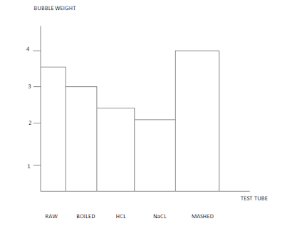
12- With a glass-marking pen mark the heigh of the bubbles. Measure it with a ruler.
13- Compare the results of the 5 test tubes.

Table with the important parts of the experiment:
RESULTS: 


 QUESTIONS:
1. How did the temperature of the potato affect the activity of catalase?  
Temperature denaturate the catalase. 
2. How did the change of the pH of the potato affect the activity of catalase?
The change of the Ph denaturate the catalase. 
3. In wich potato treatment was catalase the most active? Why do you think this was?
Mashed potato,  because we had broken the cells and the catalase was more quickly. 
4. An experiment was performed to test the effect of temperature and pH on the activity of Enzyme X. The following data was collected during the experiment: 


a) What is the optimum pH of enzyme X? 8 (maximum activity)
b) What is the optimum temperature of enzyme X? 20 (maximum activity)
c) Why do you think enzyme X has low activity at a pH of 10? Because has been denatured 
 d) Enzyme X perfoms critical life functions. Use the data above to explain why a fever of  40 degrees may be dangerous. 
 Beacuse is more lower than the maximum activity.